
BD PharmingenTM PE Mouse Anti-Human LAG-3 (CD223)
565617 · 25 Tests
BDB Bioscience · Cat: 565617
Reach 3+ verified distributors
Manufacturer product page
bdbiosciences.com/en-us/products/reagents/flow-cytometry-rea...Description
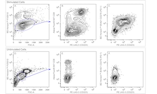
Multicolor flow cytometric analysis of LAG-3 CD223 expression on activated human peripheral blood lymphocytes. Human peripheral blood mononuclear cells were left untreated Unstimulated Cells; Bottom Panel , or were cultured Stimulated Cells; Top Panel for 3 days with plate-bound Anti-Human CD3 Cat. No. 555329; 10 mug/mL for coating and soluble Anti-Human CD28 Cat. No. 555725; 1 mug/mL antibodies, and Human Recombinant IL-2 Cat. No. 554603; 10 ng/mL . The Unstimulated and Stimulated Cells were pre-stained with BD Horizon Fixable Viability Stain 510 FVS510; Cat. No. 564406 and fixed with BD Cytofix Fixation Buffer Cat. No. 554655 . The cells were then stained with Alexa Fluor 647 Mouse Anti-Human CD8 Cat. No. 557708 , BD Horizon BV421 Mouse Anti-Human CD279 PD-1 Cat. No. 562516 , and PE Mouse Anti-Human LAG-3 CD223 Cat. No. 565616/ 565617 antibodies. Two-color flow cytometric contour plots showing the correlated expression of LAG-3 CD223 versus CD8 Plots B and E or CD279 PD-1 Plots C and F were derived from live lymphocyte-gated events ie, gated events with low level FVS510 incorporation and forward scattered-light signals FSC-A characteristic of intact lymphocytes; Plots A and D . Flow cytometric analysis was performed using a BD LSRFortessa Flow Cytometer System. Multicolor flow cytometric analysis of LAG-3 CD223 expression on activated human peripheral blood lymphocytes. Human peripheral blood mononuclear cells were left untreated Unstimulated Cells; Bottom Panel , or were cultured Stimulated Cells; Top Panel for 3 days with plate-bound Anti-Human CD3 Cat. No. 555329; 10 mug/mL for coating and soluble Anti-Human CD28 Cat. No. 555725; 1 mug/mL antibodies, and Human Recombinant IL-2 Cat. No. 554603; 10 ng/mL . The Unstimulated and Stimulated Cells were pre-stained with BD Horizon Fixable Viability Stain 510 FVS510; Cat. No. 564406 and fixed with BD Cytofix Fixation Buffer Cat. No. 554655 . The cells were then stained with Alexa Fluor 647 Mouse Anti-Human CD8 Cat. No. 557708 , BD Horizon BV421 Mouse Anti-Human CD279 PD-1 Cat. No. 562516 , and PE Mouse Anti-Human LAG-3 CD223 Cat. No. 565616/ 565617 antibodies. Two-color flow cytometric contour plots showing the correlated expression of LAG-3 CD223 versus CD8 Plots B and E or CD279 PD-1 Plots C and F were derived from live lymphocyte-gated events ie, gated events with low level FVS510 incorporation and forward scattered-light signals FSC-A characteristic of intact lymphocytes; Plots A and D . Flow cytometric analysis was performed using a BD LSRFortessa Flow Cytometer System. Multicolor flow cytometric analysis of LAG-3 CD223 expression on activated human peripheral blood lymphocytes. Human peripheral blood mononuclear cells were left untreated Unstimulated Cells; Bottom Panel , or were cultured Stimulated Cells; Top Panel for 3 days with plate-bound Anti-Human CD3 Cat. No. 555329; 10 mug/mL for coating and soluble Anti-Human CD28 Cat. No. 555725; 1 mug/mL antibodies, and Human Recombinant IL-2 Cat. No. 554603; 10 ng/mL . The Unstimulated and Stimulated Cells were pre-stained with BD Horizon Fixable Viability Stain 510 FVS510; Cat. No. 564406 and fixed with BD Cytofix Fixation Buffer Cat. No. 554655 . The cells were then stained with Alexa Fluor 647 Mouse Anti-Human CD8 Cat. No. 557708 , BD Horizon BV421 Mouse Anti-Human CD279 PD-1 Cat. No. 562516 , and PE Mouse Anti-Human LAG-3 CD223 Cat. No. 565616/ 565617 antibodies. Two-color flow cytometric contour plots showing the correlated expression of LAG-3 CD223 versus CD8 Plots B and E or CD279 PD-1 Plots C and F were derived from live lymphocyte-gated events ie, gated events with low level FVS510 incorporation and forward scattered-light signals FSC-A characteristic of intact lymphocytes; Plots A and D . Flow cytometric analysis was performed using a BD LSRFortessa Flow Cytometer System. Show More Multicolor flow cytometric analysis of LAG-3 CD223 expression on activated human peripheral blood lymphocytes. Human peripheral blood mononuclear cells were left untreated Unstimulated Cells; Bottom Panel , or were cultured Stimulated Cells; Top Panel for 3 days with plate-bound Anti-Human CD3 Cat. No. 555329; 10 mug/mL for coating and soluble Anti-Human CD28 Cat. No. 555725; 1 mug/mL antibodies, and Human Recombinant IL-2 Cat. No. 554603; 10 ng/mL . The Unstimulated and Stimulated Cells were pre-stained with BD Horizon Fixable Viability Stain 510 FVS510; Cat. No. 564406 and fixed with BD Cytofix Fixation Buffer Cat. No. 554655 . The cells were then stained with Alexa Fluor 647 Mouse Anti-Human CD8 Cat. No. 557708 , BD Horizon BV421 Mouse Anti-Human CD279 PD-1 Cat. No. 562516 , and PE Mouse Anti-Human LAG-3 CD223 Cat. No. 565616/ 565617 antibodies. Two-color flow cytometric contour plots showing the correlated expression of LAG-3 CD223 versus CD8 Plots B and E or CD279 PD-1 Plots C and F were derived from live lymphocyte-gated events ie, gated events with low level FVS510 incorporation and forward scattered-light signals FSC-A characteristic of intact lymphocytes; Plots A and D . Flow cytometric analysis was performed using a BD LSRFortessa Flow Cytometer System. Show More Multicolor flow cytometric analysis of LAG-3 CD223 expression on activated human peripheral blood lymphocytes. Human peripheral blood mononuclear cells were left untreated Unstimulated Cells; Bottom Panel , or were cultured Stimulated Cells; Top Panel for 3 days with plate-bound Anti-Human CD3 Cat. No. 555329; 10 mug/mL for coating and soluble Anti-Human CD28 Cat. No. 555725; 1 mug/mL antibodies, and Human Recombinant IL-2 Cat. No. 554603; 10 ng/mL . The Unstimulated and Stimulated Cells were pre-stained with BD Horizon Fixable Viability Stain 510 FVS510; Cat. No. 564406 and fixed with BD Cytofix Fixation Buffer Cat. No. 554655 . The cells were then stained with Alexa Fluor 647 Mouse Anti-Human CD8 Cat. No. 557708 , BD Horizon BV421 Mouse Anti-Human CD279 PD-1 Cat. No. 562516 , and PE Mouse Anti-Human LAG-3 CD223 Cat. No. 565616/ 565617 antibodies. Two-color flow cytometric contour plots showing the correlated expression of LAG-3 CD223 versus CD8 Plots B and E or CD279 PD-1 Plots C and F were derived from live lymphocyte-gated events ie, gated events with low level FVS510 incorporation and forward scattered-light signals FSC-A characteristic of intact lymphocytes; Plots A and D . Flow cytometric analysis was performed using a BD LSRFortessa Flow Cytometer System. Multicolor flow cytometric analysis of LAG-3 CD223 expression on activated human peripheral blood lymphocytes. Human peripheral blood mononuclear cells were left untreated Unstimulated Cells; Bottom Panel , or were cultured Stimulated Cells; Top Panel for 3 days with plate-bound Anti-Human CD3 Cat. No. 555329; 10 mug/mL for coating and soluble Anti-Human CD28 Cat. No. 555725; 1 mug/mL antibodies, and Human Recombinant IL-2 Cat. No. 554603; 10 ng/mL . The Unstimulated and Stimulated Cells were pre-stained with BD Horizon Fixable Viability Stain 510 FVS510; Cat. No. 564406 and fixed with BD Cytofix Fixation Buffer Cat. No. 554655 . The cells were then stained with Alexa Fluor 647 Mouse Anti-Human CD8 Cat. No. 557708 , BD Horizon BV421 Mouse Anti-Human CD279 PD-1 Cat. No. 562516 , and PE Mouse Anti-Human LAG-3 CD223 Cat. No. 565616/ 565617 antibodies. Two-color flow cytometric contour plots showing the correlated expression of LAG-3 CD223 versus CD8 Plots B and E or CD279 PD-1 Plots C and F were derived from live lymphocyte-gated events ie, gated events with low level FVS510 incorporation and forward scattered-light signals FSC-A characteristic of intact lymphocytes; Plots A and D . Flow cytometric analysis was performed using a BD LSRFortessa Flow Cytometer System.