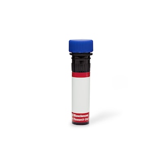
BD PharmingenTM 647 EdU Click Proliferation Kit
565456 ·
BDB Bioscience · Cat: 565456
Reach 3+ verified distributors
Manufacturer product page
bdbiosciences.com/en-us/products/reagents/flow-cytometry-rea...
Description
Figure 1. Cell Cycle Analysis of Proliferating HeLa cells. HeLa cells were pulsed with 10 muM EdU for 1 hour, and then fixed, permeabilized, and stained with Eterneon Red azide azide and BD Pharmingen DAPI Solution Cat. No. 564907 according to the recommended assay procedure. Histograms were derived from gated events based on light scattering characteristics of intact HeLa cells. Flow cytometric analysis was performed using a BD LSRFortessa Flow Cytometry System. Figure 2. Fluorescent Staining of HeLa Cells for EdU Incorporation and Histone H3 pS28 . HeLa cells were seeded in a 96-well imaging plate at 15,000 cells per well. After overnight incubation, cells were pulsed with 10 muM EdU for 1 hour, and then fixed, permeabilized, and stained with Eterneon Red azide pseudo-colored red according to the Recommended Assay Procedure. Cells were washed twice and stained with Alexa Fluor 488 Rat Anti-Histone H3 pS28 pseudo-colored green, Cat. No. 558610 . Nuclei were counterstained with DAPI pseudo-colored blue . Eterneon Red appears magenta when colocalized with DAPI, and Alexa Fluor 488 Rat Anti-Histone H3 pS28 appears cyan when colocalized with DAPI. Images were captured on a BD Pathway 435 Bioimager System with a 20X objective and merged using BD Attovision software. Figure 1. Cell Cycle Analysis of Proliferating HeLa cells. HeLa cells were pulsed with 10 muM EdU for 1 hour, and then fixed, permeabilized, and stained with Eterneon Red azide azide and BD Pharmingen DAPI Solution Cat. No. 564907 according to the recommended assay procedure. Histograms were derived from gated events based on light scattering characteristics of intact HeLa cells. Flow cytometric analysis was performed using a BD LSRFortessa Flow Cytometry System. Figure 2. Fluorescent Staining of HeLa Cells for EdU Incorporation and Histone H3 pS28 . HeLa cells were seeded in a 96-well imaging plate at 15,000 cells per well. After overnight incubation, cells were pulsed with 10 muM EdU for 1 hour, and then fixed, permeabilized, and stained with Eterneon Red azide pseudo-colored red according to the Recommended Assay Procedure. Cells were washed twice and stained with Alexa Fluor 488 Rat Anti-Histone H3 pS28 pseudo-colored green, Cat. No. 558610 . Nuclei were counterstained with DAPI pseudo-colored blue . Eterneon Red appears magenta when colocalized with DAPI, and Alexa Fluor 488 Rat Anti-Histone H3 pS28 appears cyan when colocalized with DAPI. Images were captured on a BD Pathway 435 Bioimager System with a 20X objective and merged using BD Attovision software. Show More Figure 1. Cell Cycle Analysis of Proliferating HeLa cells. HeLa cells were pulsed with 10 muM EdU for 1 hour, and then fixed, permeabilized, and stained with Eterneon Red azide azide and BD Pharmingen DAPI Solution Cat. No. 564907 according to the recommended assay procedure. Histograms were derived from gated events based on light scattering characteristics of intact HeLa cells. Flow cytometric analysis was performed using a BD LSRFortessa Flow Cytometry System. Figure 2. Fluorescent Staining of HeLa Cells for EdU Incorporation and Histone H3 pS28 . HeLa cells were seeded in a 96-well imaging plate at 15,000 cells per well. After overnight incubation, cells were pulsed with 10 muM EdU for 1 hour, and then fixed, permeabilized, and stained with Eterneon Red azide pseudo-colored red according to the Recommended Assay Procedure. Cells were washed twice and stained with Alexa Fluor 488 Rat Anti-Histone H3 pS28 pseudo-colored green, Cat. No. 558610 . Nuclei were counterstained with DAPI pseudo-colored blue . Eterneon Red appears magenta when colocalized with DAPI, and Alexa Fluor 488 Rat Anti-Histone H3 pS28 appears cyan when colocalized with DAPI. Images were captured on a BD Pathway 435 Bioimager System with a 20X objective and merged using BD Attovision software.
More from BDB Bioscience